Development, image interpretation, clinical experience, and applications of optical coherence tomography in neurointerventional surgery
J NeuroIntervent Surg:
20 February 2026.
Our proprietary AI enabled neuro Optical Coherence Tomography (nOCT) platform is bringing high resolution intravascular imaging inside the brain for the first time.
With our platform’s integrated AI capabilities, we are collaborating with our clinical partners to enhance neurovascular diagnostic capabilities to help accelerate development of new therapies & ultimately improve patient care worldwide.
![]()
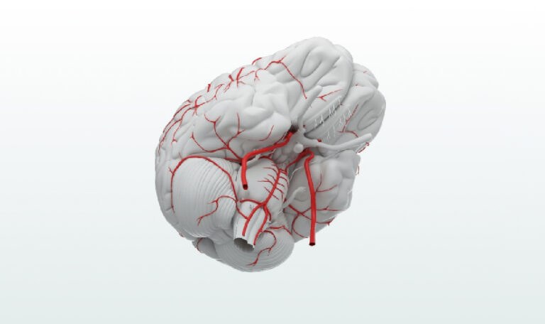
neuro Optical Coherence Tomography
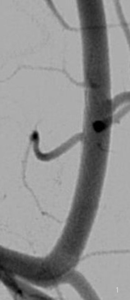
Enabling you to see past the boundaries of the arterial lumen, through the arterial wall and even into the perivascular space.
New knowledge, new patient insights.
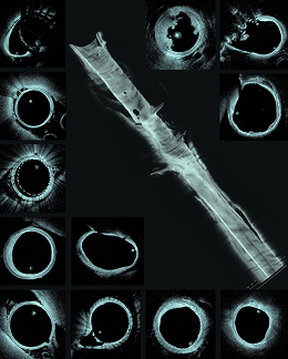
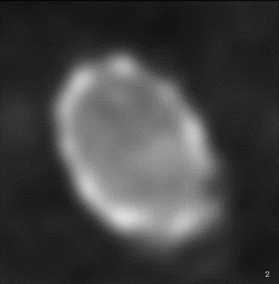
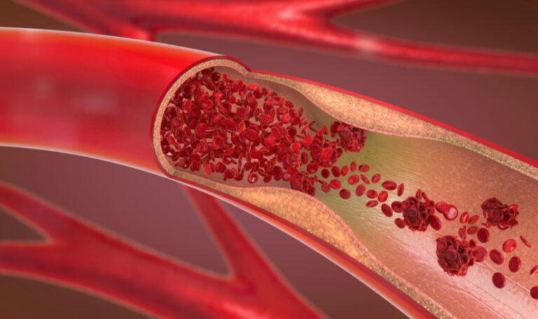
Intravascular optical microscopy provided by nOCT can offer unique insights into the treatment of brain aneurysms, ischemic stroke, and atherosclerotic intracranial disease by revealing details of the vessel wall and devices that remain hidden from non-invasive imaging modalities.
Neurointerventionalists will be able to routinely visualize anatomic structures, diseases, and therapies, to understand their etiology and interactions at a level previously unattainable.

Revolutions in neurointerventional imaging, from the discovery of x-rays over 120 years ago, to 3-dimensional imaging in the angiography suite 20 years ago, occur roughly once in a generation. Direct imaging of the pathology and its relationship to devices transforms treatment decisions, and the fundamental understanding of cerebrovascular pathology.
- Matthew Gounis, Ph.D.
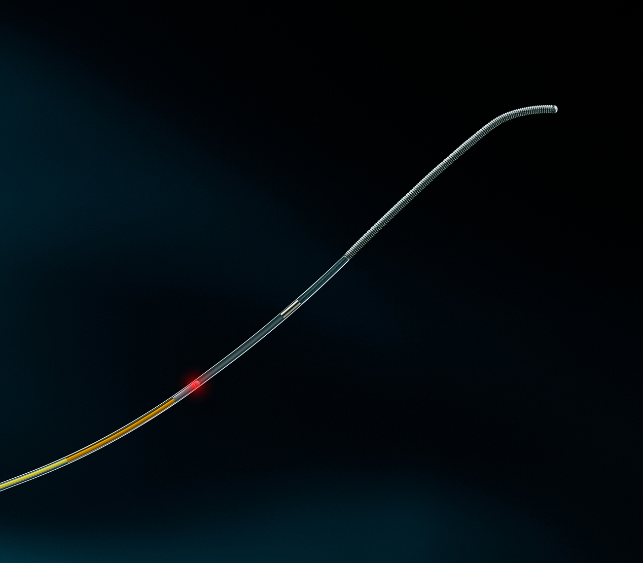
Designed to navigate the highly tortuous neurovascular anatomy
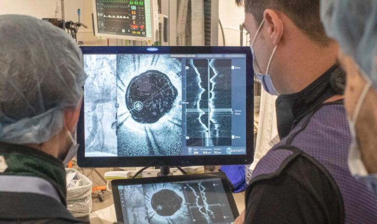
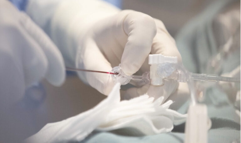
Using standard neurovascular workflow, the Spryte nOCT imaging probe brings a micro-optical fiber into the brain’s arteries, allowing for imaging from the inside out. The imaging platform includes an AI-powered console, an optical module, and a fiber-optic, neuroendovascular probe specifically designed to navigate the small, tortuous vasculature of the brain.

The Spryte nOCT technology is an imaging probe that actually works like a wire, and it navigated perfectly through the brain vessels. The system and imaging probes performed very well, integrating with our workflow seamlessly, and provided us with important information that we cannot obtain with any other technology – very impressive.
- Vitor Mendes Pereira, M.D., MSc
nOCT images vessels, disease and other important structures at >10x the resolution of today’s imaging systems and improving clinicians’ understanding of neurovascular disease.
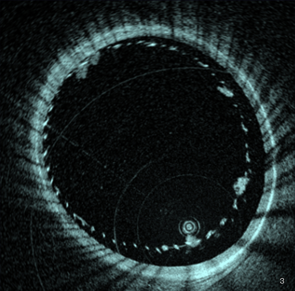
Clot sitting on the struts of the flow diverting stent, not visible with traditional non-invasive imaging like angiography and Cone beam CT.
J NeuroIntervent Surg:
20 February 2026.
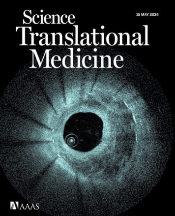
Science Translational Medicine,
15 May 2024, Vol 16, Issue 747

Because of neuro OCT, we will now be able, for the first time, to analyze and better understand different pathologies and the results of our neurointerventional therapies. It is a real game changer in the cerebrovascular field.
- Pedro Lylyk, MD
Dr. Vitor Mendes Pereira, St Michael’s Hospital Toronto
Spryte Medical Announces First Participants Enrolled in the INSYTE Trial at Baptist Health Jacksonville
Globe Newswire (Press Release), March 3, 2026
Spryte Medical Receives FDA IDE Approval to Initiate “INSYTE” US Pivotal Trial evaluating the nOCT Imaging System during Brain Aneurysm Treatment
Globe Newswire (Press Release), November 19, 2025
Spryte Medical Receives Breakthrough Device Designation from FDA for Revolutionary neuro Optical Coherence Tomography (nOCT) Technology
Business Wire (Press Release), July 22, 2024
"Snake-like" Probe Images Arteries from Within - IEEE Spectrum
IEEE Spectrum, May 15, 2024
Spryte Medical Announces First Human Use of Novel Intravascular Brain Imaging Technology, Published in Science Translational Medicine | Business Wire
Business Wire (Press Release), May 15, 2024
Study finds intravascular imaging effective for imaging the brain - Medical Device Network
Medical Device Network, May 14, 2024
Spryte publishes first-in-human study of novel intravascular brain imaging technology
NeuroNews, May 24, 2024
Miniaturized Snake-Like Probe Images Cerebral Arteries From Within
HospiMedica, May 20, 2024

Spryte Medical Announces First Human Use of Novel Intravascular Brain Imaging Technology, Published in Science Translational Medicine
Asian Hospital & Healthcare Management, May 16, 2024

Miniaturized optical coherence tomography imaging probe takes pictures inside cerebral arteries
Medical Xpress, May 16, 2024
To spot an incipient stroke, tiny brain probe acts ‘like a microscope’
STAT, May 15, 2024
St. Michael’s Performs Another World-First Procedure
St Michael's Foundation
Spryte Medical’s mission is to fundamentally transform and improve stroke patient care. Our proprietary AI enabled neuro Optical Coherence Tomography (nOCT) platform is bringing high resolution intravascular imaging inside the brain for the first time. With this approach we can image vessels, disease, and other important structures at >10x the resolution of today’s imaging systems, thus improving clinicians understanding of stroke and other neurovascular disease. In collaboration with our clinical partners we are working to accelerate development of new therapies, enhance procedural techniques and improve patient care worldwide.
Thank You